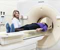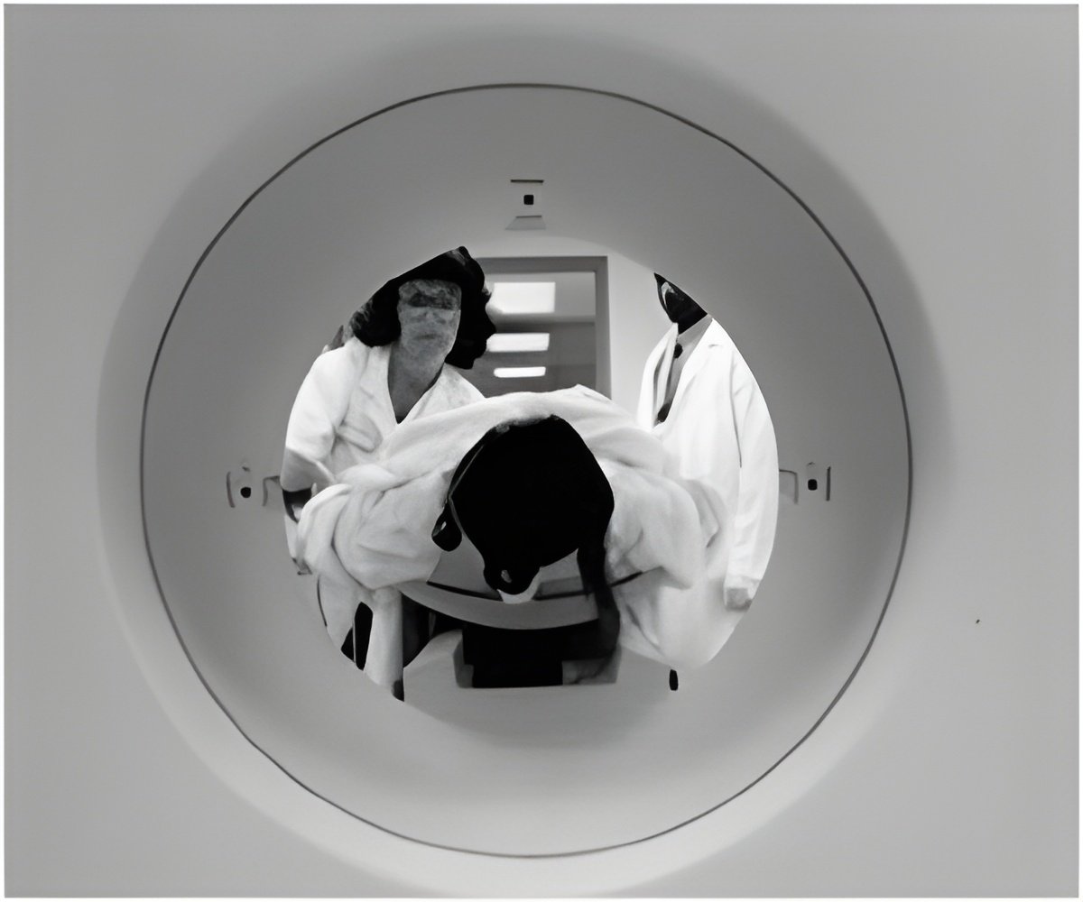
Conventional medical imaging tools – including X-ray, ultrasound, CT and MRI – detect disease by finding the anatomy, or shape and size, of the abnormality. When using these tools to screen for cancer, a tumor must be large enough to be detected, and if found, a surgical biopsy is generally required to determine if it is benign or malignant.
Duke researchers are working to develop imaging technologies to detect disease in its earliest stages, much before the tumors grow large enough to be detected using conventional methods. Two imaging techniques they are researching are Neutron Stimulated Emission Computed Tomography and Gamma Stimulated Emission Computed Tomography.
Research has shown that many tumors have an out-of-balance concentration of trace-level elements naturally found in the body, such as aluminum and rubidium. These elements stray from their normal concentration levels at the earliest stages of tumor growth, potentially providing an early signal of disease.
The neutron and gamma imaging methods measure the concentrations of elements in the body, determining molecular properties without the need for a biopsy or injection of contrast media. The goal is for these tests to be able to distinguish between benign and malignant lesions, as well as healthy tissue.
Advertisement

