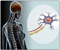
The new study is based on a group of 40 multiple sclerosis (MS) patients, who used a process called optical coherence tomography (OCT) to scan the layers of nerve fibers of the retina in the back of the eye, which become the optic nerve.
OCT, which uses a desktop machine similar to a slit-lamp, is simple and painless. The retinal nerve fiber layer is the one part of the brain where nerve cells are not covered with the fat and protein sheathing called myelin, making this assessment specific for nerve damage as opposed to brain MRI changes, which reflect an array of different types of tissue processes in the brain.For the study, results of the scans were calibrated using accepted norms for retinal fiber thickness and then compared to an MRI of each of the patient’s brains - also calibrated using accepted norms.
After analysis, researchers found a correlation coefficient of 0.46, after accounting for age differences. Correlation coefficients represent how closely two variables are related -- in this case MRI of the brain and OCT scans.
Correlation coefficients range from -1 (a perfect opposing correlation) through 0 (no correlation) to +1 (a perfect positive correlation). In a subset of patients with relapsing remitting MS, the most common form of the disease, the correlation coefficient jumped to 0.69, suggesting an even stronger association between the retinal measurement and brain atrophy.
“This is an encouraging result,” said Johns Hopkins neurologist Peter Calabresi, M.D., lead author of the study.
“MRI is an imperfect tool that measures the result of many types of tissue loss rather than specifically nerve damage itself. With OCT we can see exactly how healthy these nerves are, potentially in advance of other symptoms,” he added.
Advertisement
OCT scans look directly at the thickness, and therefore health, of the optic nerve, which is affected early on in the disease, often before the patient suffers permanent brain damage, he added.
Advertisement
Calabresi said that his next step would be to look at changes in the fiber layer thickness in 100 patients over a period of three years.
The new study is published in the October 2007 issue of Neurology
Source-Eurekalert
SPH/V


