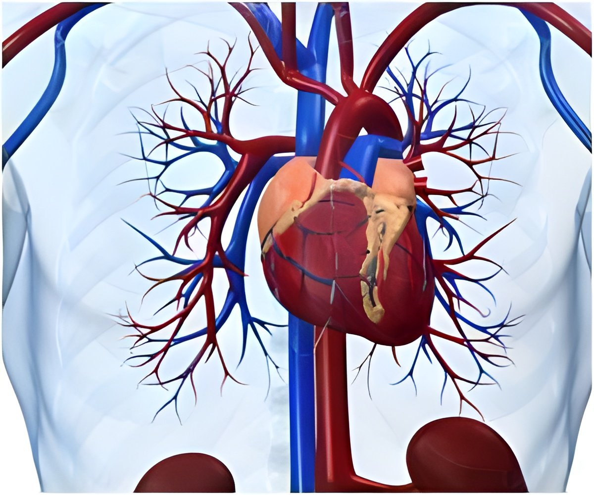
Consultant congenital cardiologist Dr Joseph Vettukattil pioneered the technique's development at Southampton General Hospital, Hampshire, to identify heart abnormalities present from birth.
"You can chop the heart into small pieces and see what is wrong and exactly where it is wrong on the screen," The Sydney Morning Herald quoted him, as describing the method.
He continued: "By using MPR, because you are slicing and seeing it in three different planes, you can get a clear understanding of a patient - especially in a child whose heart is congenitally malformed.
"The most important aspect is the operator's ability to slice the dynamic cardiac structures in infinite sections through all the three dimensions, which was not possible before we developed MPR 3D echocardiography."
Traditionally, diagnosis of heart defects has been made using 2D scans and invasive operations.
Advertisement
"Now, though, we are able to visualise even more than a surgeon can during an operation, minimising the need for additional and invasive assessments."
Advertisement


