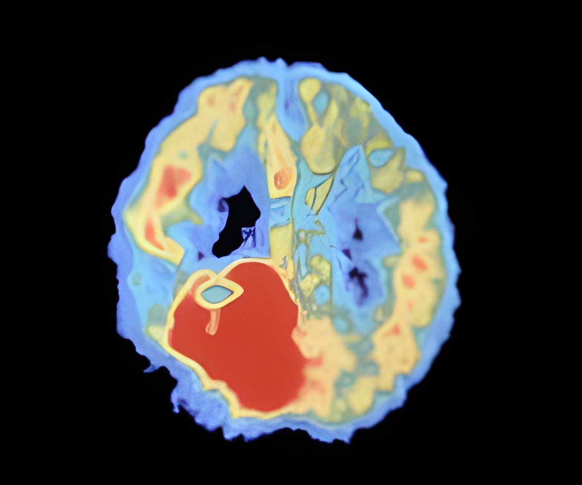
The team used an entirely new imaging method called 'functional electrical impedance tomography by evoked response' (fEITER *), which aids high speed imaging and monitoring of electrical activity deep within the brain and helps researchers measure brain function.
The machine itself is a portable, lightweight monitor, which can fit on a small trolley. It carries out an 'electronic scan' in 100 times of a second.
The speed of the response of fEITER is such that the evoked response of the brain to external stimuli, such as an anesthetic drug, can be captured in rapid succession as different parts of the brain respond, thus tracking the brain's processing activity.
"We have looked at 20 healthy volunteers and are now looking at 20 anaesthetised patients scheduled for surgery. We are able to see 3-D images of the brain's conductivity change, and those where the patient is becoming anaesthetised are most interesting," said Prof Pollard.
"We have been able to see a real time loss of consciousness in anatomically distinct regions of the brain for the first time. We are currently working on trying to interpret the changes that we have observed," he added.
Advertisement


