AIRWAY CONTROL & MAINTENANCE
Common Bed Side Procedure No. 7
Section Editor: Prof. T.K. Partha Sarathy
Introduction
Warning
A - Opening the airway : Jaw Thrust Method
B - Opening the airway: Chin lift method
C - Oropharyngeal airway
D - Nasopharyngeal airway
Endotracheal intubation
Orotracheal intubation
Nasotracheal Intubation
Introduction
Life Saving measures in any emergency situation is described under the acronym ABCD.
A stands for airway. Unless the airway is patent breathing (B) will not be adequate or possible and this will undoubtedly lead to cardiac (C) circulatory problems. If the airway problem is taken care of, most of the emergency situations could be avoided.
The problems that interfere with airway is obstruction to the passage anywhere from the nostrils down to the larynx and trachea by any one of the processes described below. It causes limitation of air entry into the lungs resulting in hypoxia leading to death.The airway is normally protected by the patients ability to swallow or cough out and clear nasopharyngeal, oropharyngeal and tracheal secretions. However, in an unconscious patient when these reflexes do not function well, these protective mechanisms will not be available.Obstruction to airway is caused by :
1. Tongue falling backwards obstructing the oropharynx in an unconscious patient
2. Secretions that collect in the oropharynx in an unconscious patient who is not able to clear it
3. The stomach contents being aspirated into larynx and trachea and filling up the oropharynx
4. Facial injuries resulting in nasopharyngeal trauma causing continuous oozing of blood and clots causing obstruction to the nasopharynx and oropharynx.
5. Foreign bodies inadvertently aspirated into larynx and trachea resulting in acute obstruction (dentures)
6. Anesthetic agents and toxic substances ingested or inhaled causing the loss of gag reflux.
7. Any unconscious status - coma due to disease or drugs - anesthesia, seizure disorders - Electro convulsive therapy etc.
Irrespective of the cause of obstruction the effect is the same. The family physician and the first contact person should be able to do a few basic preliminary procedures that would prevent disastrous results due to airway obstruction.
Warning
In any patient who has sustained multiple trauma or had a fall from heights or has head injury, it is always better to suspect injury to the cervical spine and he/she has to be handled carefully in such a manner that the neck is not manipulated or extended and appropriate neck support provided.
Airway management
The first step, which most often helps the patient in managing airway, is to maintain him in a supine posture. In this position the whole body could be tilted to one side like a log to provide drainage from the mouth to gravitate out. When there is no suspicion of neck injury this could be accomplished just by turning the head to one side.
The next step is to make sure that all foreign bodies and secretions are removed from the back of the throat. The jaw can be lifted with a thumb inside the oral cavity pressing on the tongue and the other fingers protecting the chin with the left hand.
The right hand index and middle fingers (gloved) is swept inside the mouth and with a swooping movement foreign bodies and secretions in the oropharynx are brought out. This is followed by adequate suctioning of the oropharynx to clear of secretions, aspirates from the stomach, foreign bodies etc. If the tongue is flopping backwards applying a tongue forceps to the anterior tongue this can be drawn forward to provide direct airway without obstruction.
(Arrows point to airway obstruction due to tongue falling backwards).
A - Opening the airway : Jaw Thrust Method
|
|
|

B - Opening the airway: Chin lift method
|
|
Fingers of one hand placed under the mandible and gently lift the chin forwards. The lower lip is depressed with the thumb to open the mouth. |

| Note: Head tilt as shown above will help improve airway. But hyperextension or flexion of neck must be avoided if cervical spine injury is suspected or has not been ruled out. |
|
|
Requirement: |

Procedure:
D - Nasopharyngeal airway
Ideal when patient is somewhat conscious - better tolerated than oropharyngeal.
|
|
Requirements: |

Procedure:
Definitive Airway Control and Management
Endotracheal intubation: Consider endotracheal intubation when definitive airway is needed under the following circumstances:
a. If patients airway is obstructed inspite of the placement of oropharyngeal/nasopharyngeal airway and ventilation.
b. Patient is not breathing (apnoea)
c. Inadequate air exchange with or without other problems like pneumothorax, hemothorax, pericardial tamponade etc. all would require immediate intervention.
d. To prevent aspiration of stomach contents into the trachea in unconscious patients
e. Where inhalation burns, status epileptics, head trauma and facial fractures are suspected.
Endotracheal intubation and ventilation is a safe and reliable method of oxygenation. Endotracheal intubation is a skill that can be learnt with ease and confidence and undoubtedly is a Life Saving Measure that every family practitioner should handle competently. Endotracheal intubation can be performed through the oral cavity (orotrachea) or through the Nasal cavity (Nasotrachea). Both these methods are simple skills that require all paramedics, nurses, primary care physicians and emergency personnel should learn.(We recommend that during contact sessions DFH students practice these skills with a manikin).
Learning skills:
- Basic anatomy of the nasooropharynx, glottis and trachea should be understood
- The glottis opening should be easily recognized
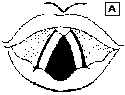 |
Please refer to the Anatomical diagram A and identify the vallecula, vocal cords, epiglottis and glottic opening. |
|
|
On the diagram B mark tongue, pharynx, vallecula, epiglottis, glottic opening and the trachea. |
|
|
This simple exercise will familiarize with the basic anatomical knowledge that is required for endotracheal intubation. |


Laryngoscope has a handle which contains batteries and a blade with a bulb for light source. The blade can be either a straight one or a curved one. The handles and blades come in different sizes to suite different age groups. It is very important that every family practitioner has in possession the following
items:
1. Adult laryngoscope with at least one curved blade, one straight blade, spare bulbs and batteries
2. One similar paediatric set
3. Endotracheal tubes of different sizes
4.Ambu bags.
|
|
Figure D shows endotracheal tube and they come in different sizes. The modern endotracheal tube has a inflatable cuff at the end and through a separate portal the cuff can be inflated by injecting a few ccs or air. By utilizing the cuffed endotracheal tube the following are accomplished: |
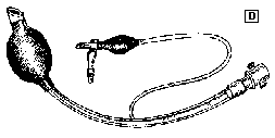
a. It is possible to prevent aspiration of stomach contents totally by snugly inflating the cuff
b. The air filled sac is less traumatizing to the mucosa of the trachea than the rough tube itself and less likely to scrip out.
c. If a patient has to be ventilated air leak will not take place.
We recommend that Family Practitioners use cuffed endotracheal tubes. These are disposable ones and are easily available.
Orotracheal intubation:
Requirements:
a. Laryngoscope
b. Endotracheal tubes (stylet included)
c. Suction machine
d. 10 cc Syringe
e. Ambu bag / ventilator
f. Stethoscope
Position: Endotracheal intubation in emergencies is done with a patient in recumbent posture and where neck injury is suspected, an assistant to hold the neck in neutral position without hyperextension or flexion is recommended.
Procedure:
| Note: Only way one to be sure that the tube is not in the esophagus and that it is well in trachea is by a careful examination performed on both sides of the chest to watch for movement of the chest as well as breath sounds heard on auscultation. |
Endotracheal intubation has to be accomplished promptly and quickly without wasting time and it is better to have everything ready before starting the procedure while making sure that the patient is being oxygenated and ventilated. In an apneic patient this has to be accomplished with precision and speed.It is better not to make more than one or two trials of introducing the tube and if it is not successful, try a few seconds later after adequately oxygenating and ventilating with a face mask and ambu bag. Repeated traumatizing efforts without giving breaks with ventilatory support should not be attempted. After the placement of endotracheal tube if there is evidence of thick secretions or gastric aspirate this has to be suctioned out. Frequent, continuous suction without oxygenation and ventilation in between should be avoided. Intermittent suction after adequate ventilation is the procedure of choice when necessary. Where facilities exist O2 saturation level could be documented using pulse oximeter.
| Note: At times it becomes difficult to expose the glottis utilizing a curved laryngoscopic blade. In such cases it may be worthwhile making the next attempt with a straight blade. |
|
|
Advantage: When curved blade is used, the epiglottis is pulled upwards with the tip of the blade in the vallecula (E), |
|
|
where as with the straight blade it is placed on the epiglottis which is elevated to expose the glottis (F). |


The use of a stylet inside the endotracheal tube may come in handy to make the passage of the tube through the glottis easier and also to contour the tube curvature as needed. If a stylet is used it must be withdrawn as soon as optimal entry of the tube in the trachea is obtained.
Nasotracheal Intubation
Under most circumstances orotracheal intubation is adequate and is quicker to accomplish. In select circumstances, and in particular, in a semi-alert patient with head injury it may be beneficial to use nasotracheal intubation. Once placed properly it is tolerated and retained by the patients much better and is unlikely to be coughed up, chewed or spit.
Requirements:
The requirements for nasotracheal intubation is essentially the same. Xylocaine Jelly is recommended for the lubrication of the nostril as well as the tube.
Contra indications:
1. Nasal and facial bone fracture
2. In apneic patients nasotracheal intubation would require to listen to breath sounds and in apneic patients that is not possible.
3. In patients with basal skull fracture and CSF rhinorrhea.
Procedure:
However, it is always better to confirm the position of the endotracheal tube with a chest x-ray taken as soon as possible.
Although some what difficult to perform nasotracheal intubation skill comes in handy in select patients and have distinct advantages.


Comments
I'm a 23 year old male,it was only today that I came to know about my paraphimosis. It happened because of my own mistakes,at that time I my foreskin was swollen badly,there was pain. But I didn't tell parents or consulted a doctor because I was ashamed of myself. I know it was a wrong choice. However after 15 days pain was gone,and everything was normal except that my foreskin wont come back on its usual place,and it was stuck behind the glan forming oedema. It's been 6 years, I can do all the normal thing. But my oedema is still there. I want to know if it can cause any problem in future?
what can i do as a nurse when the female urethral meatus during cathetherization is invisible.
posted by shadrack kipkorir , from kenya
what are the problems associated with catheterization
I have undergone dorsal slit operation 20 days ago,now the swollen in completely cured however the cut on the foreskin has not healed till now, pls suggest how long will it take to completely heal so that i can go for regular intercourse.RAHUL.
I would like ask about this. I have pain in left mandibular if I open my mouth so I just open my mouth not normal. This condition has happened for 2 weeks. what should I do?
thank you..:0